Rish Academy

Gabapentin Ruined My Life rish academy

Gabapentin Ruined My Life

Newsletter Gabapentin Ruined My Life Gabapentin Ruined My Life: The Dark Side of a Popular Medication In the realm of pharmaceuticals, there are drugs that bring relief, hope, and improved quality of life. However, every once in a while, a medication emerges that carries unforeseen consequences, leaving a trail of despair in its wake. One such drug is gabapentin, a medication that was initially developed to treat epilepsy but is now commonly prescribed for a range of conditions, including chronic pain, neuropathy, and anxiety disorders. While gabapentin has helped many individuals manage their symptoms effectively, there is a growing concern over its potential for abuse, dependence, and debilitating side effects. In this blog article, we delve into the experiences of those who claim that gabapentin ruined their lives.   Rish Academy’s Pharmacology Flashcards eBook Click the button below to download Rish Academy’s Pharmacology Flashcards eBook. The book has 15 chapters and 122 Pages.  Download Now How Gabapentin works? Gabapentin works by affecting certain neurotransmitters in the brain, specifically gamma-aminobutyric acid (GABA). GABA is an inhibitory neurotransmitter that helps regulate the transmission of signals between nerve cells. By modulating GABA activity, gabapentin exerts its therapeutic effects. While the exact mechanism of action of gabapentin is not fully understood, it is believed to have multiple mechanisms that contribute to its effects: 1. Binding to the α2δ subunit of voltage-gated calcium channels: Gabapentin binds to a specific subunit (α2δ) of voltage-gated calcium channels in the brain. By doing so, it inhibits the entry of calcium ions into nerve cells, which reduces the release of certain excitatory neurotransmitters. This modulation of calcium channels helps regulate the transmission of pain signals and can contribute to the analgesic effects of gabapentin. 2. Increasing GABA synthesis: Gabapentin has been shown to increase the synthesis of GABA, the inhibitory neurotransmitter. This can enhance GABAergic signaling in the brain, leading to a calming and sedative effect. 3. Modulating glutamate release: Gabapentin may also modulate the release of glutamate, an excitatory neurotransmitter. It is thought to reduce excessive glutamate release, which can help dampen nerve excitability and reduce seizures or neuropathic pain. Over the years, gabapentin has gained popularity due to its perceived effectiveness in treating various conditions beyond epilepsy. Doctors have prescribed it for off-label uses such as fibromyalgia, restless leg syndrome, migraines, and even as an adjunct treatment for opioid withdrawal. However, as its use expanded, so did reports of adverse effects and misuse. As a medical community, it is imperative that we listen to the voices of those affected and conduct further research to gain a comprehensive understanding of gabapentin’s long-term effects. By doing so, we can improve patient outcomes, mitigate potential harm, and ensure that individuals receive appropriate and safe treatments tailored to their specific needs. Click the button below to download Rish Academy’s Pharmacology Flashcards eBook. The book has 15 chapters and 122 Pages.  Download Now What are the dangers of gabapentin? Gabapentin, like any medication, carries certain risks and potential dangers. While it is important to note that not everyone will experience these effects, it is crucial to be aware of the possible risks associated with gabapentin use. Here are some of the dangers and potential adverse effects: 1. Dependency and Abuse: Although gabapentin is not classified as a controlled substance in many countries, there is increasing evidence of its potential for abuse and dependence. Some individuals may develop a psychological or physical dependence on the medication, leading to difficulties in discontinuing its use without experiencing withdrawal symptoms. 2. Increased Risk of Suicidal Thoughts and Behavior: Gabapentin has been associated with an increased risk of suicidal thoughts and behaviors, particularly in individuals with pre-existing mental health conditions such as depression or anxiety. It is crucial to monitor patients closely, especially when starting or changing the dosage of gabapentin. 3. Cognitive Impairment: Many individuals who take gabapentin for an extended period report cognitive side effects, such as memory problems, difficulty concentrating, and impaired thinking. These effects can impact daily functioning, work performance, and overall quality of life. 4. Sedation and Drowsiness: Gabapentin can cause drowsiness, dizziness, and sedation, particularly when taken at higher doses or in combination with other medications that have similar effects. This can increase the risk of accidents, falls, and impaired judgment. 5. Increased Risk of Respiratory Depression: In rare cases, gabapentin has been associated with respiratory depression, particularly when combined with other central nervous system depressants, such as opioids or benzodiazepines. This can be life-threatening and requires immediate medical attention. 6. Emotional Instability and Mood Changes: Some individuals may experience emotional instability, mood swings, increased anxiety, or depression as a result of gabapentin use. These effects can be distressing and may require close monitoring and adjustment of the treatment plan. 7. Physical Side Effects: Gabapentin can cause a range of physical side effects, including fatigue, weakness, dizziness, coordination problems, blurred vision, and weight gain. These side effects can vary in severity and may affect individuals differently. It is important to note that these dangers and side effects are not exhaustive, and individual experiences may vary. It is essential to discuss any concerns or potential risks with a healthcare professional before starting or discontinuing gabapentin treatment. Healthcare providers can provide personalized advice and closely monitor patients to ensure their safety and well-being during gabapentin therapy. Click the button below to Download 570+ High-Yield Presentations in Emergencies, Orthopedics, Gynaecology & Obstetrics, Surgery, and Clinical Medicine Get LIFETIME Access to 570+ Medical Presenations What is the biggest side effect of Gabapentin? While gabapentin can cause various side effects, one of the most commonly reported and significant side effects is drowsiness or sedation. Many individuals who take gabapentin experience increased sleepiness or a feeling of being excessively tired. This side effect can range from mild drowsiness to profound sedation, depending on the dosage and individual sensitivity to the medication. Drowsiness caused by gabapentin can affect daily activities, work performance, and overall quality of life. It may impair cognitive function, making it difficult to concentrate,

Read More

Mitochondrial-Inheritance-Examples-rish-academy-img

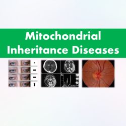
Examples of Mitochondrial Inheritance Diseases

Newsletter Mitochondrial Inheritance Examples Examples of Mitochondrial Inheritance Diseases Mitochondrial inheritance is a fascinating aspect of genetics that determines how certain genetic traits are passed down from generation to generation. Unlike nuclear DNA, which is inherited from both parents, mitochondrial DNA (mtDNA) is primarily inherited from the mother. This unique inheritance pattern offers valuable insights into the evolution, diagnosis, and treatment of various mitochondrial disorders. In this article, we will explore mitochondrial inheritance in detail, providing you with comprehensive examples that highlight its significance in medical research. Click the button below to Download 570+ High-Yield Presentations in Emergencies, Orthopedics, Gynaecology & Obstetrics, Surgery, and Clinical Medicine Get LIFETIME Access to 570+ Medical Presenations Mitochondria and Their Role in Cellular Energy Production Mitochondria are small, specialized structures found within cells that are often referred to as the “powerhouses” due to their vital role in energy production. They convert nutrients into adenosine triphosphate (ATP), the cellular fuel that powers essential biological processes. Each mitochondrion contains its own circular DNA molecule, mtDNA, which encodes crucial genes involved in energy production and other cellular functions. Maternal Inheritance of mtDNA One of the remarkable characteristics of mtDNA is its predominantly maternal inheritance pattern. During fertilization, the sperm contributes only nuclear DNA to the resulting embryo, while the egg provides both nuclear DNA and mitochondria. As a result, the mtDNA from the sperm is largely eliminated shortly after fertilization, leaving the maternal mtDNA to populate the developing embryo. Heteroplasmy and Homoplasmy Mitochondrial inheritance introduces the concepts of heteroplasmy and homoplasmy. Heteroplasmy refers to the presence of more than one type of mtDNA within an individual’s cells, while homoplasmy denotes the presence of a single type of mtDNA. Heteroplasmy can arise due to various factors, such as mutations or a mixture of normal and mutated mtDNA. The proportions of different mtDNA types in heteroplasmic individuals can vary among tissues and may change over time. Click the button below to Download 570+ High-Yield Presentations in Emergencies, Orthopedics, Gynaecology & Obstetrics, Surgery, and Clinical Medicine Get LIFETIME Access to 570+ Medical Presenations Mitochondrial Inheritance Examples Here are a few examples of diseases and conditions that exhibit mitochondrial inheritance: 1. Leber’s Hereditary Optic Neuropathy (LHON): LHON is a mitochondrial disorder that primarily affects the optic nerve, leading to sudden and severe vision loss. It is caused by specific mutations in the mitochondrial DNA (mtDNA). LHON follows a maternal inheritance pattern, meaning it is primarily passed from an affected mother to her children. While both males and females can inherit the condition, males are more likely to develop symptoms. This inheritance pattern occurs because the sperm does not contribute mitochondria during fertilization, and thus, only the mother’s mtDNA is passed on to the offspring. Optic nerve with peripapillary telangiectasias and pseudoedematous appearance. 2. Mitochondrial Encephalopathy, Lactic Acidosis, and Stroke-like Episodes (MELAS) MELAS is a mitochondrial disorder characterized by recurrent stroke-like episodes, muscle weakness, and lactic acidosis. It is caused by mutations in mtDNA. MELAS follows a maternal inheritance pattern, with affected mothers transmitting the condition to their children. The severity and age of onset can vary among individuals, but the underlying genetic mutation is always present in the mitochondria inherited from the mother. Basal ganglia calcification, cerebellar atrophy, increased lactate; a CT image of a person diagnosed with MELAS Click the button below to Download 570+ High-Yield Presentations in Emergencies, Orthopedics, Gynaecology & Obstetrics, Surgery, and Clinical Medicine Get LIFETIME Access to 570+ Medical Presenations 3. Kearns-Sayre Syndrome (KSS) KSS is a rare mitochondrial disorder that affects multiple organ systems, including the eyes, heart, and muscles. Symptoms may include progressive external ophthalmoplegia (eye muscle weakness), cardiac conduction defects, and muscle weakness. KSS is typically caused by large-scale deletions or rearrangements in mtDNA. Similar to other mitochondrial disorders, KSS follows a maternal inheritance pattern, with affected mothers passing the mutated mtDNA to their children. Bilateral ptosis and external ophthalmoplegia. Top: patient looking straight ahead. Below: patient is being asked to look in the direction of the arrow in each case. Restriction of eye movements in each direction is demonstrated. 4. Mitochondrial Diabetes Mitochondrial diabetes refers to a form of diabetes that is associated with mitochondrial dysfunction caused by mutations in mtDNA. This type of diabetes often presents with features such as early age of onset, neurologic abnormalities, hearing loss, and other organ-specific manifestations. The inheritance pattern follows the maternal lineage, meaning affected mothers can pass the condition to their children. Relationship between mitochondrial dysfunction and type 2 diabetes. 5. Leigh Syndrome Leigh syndrome is a severe neurological disorder that usually presents in infancy or early childhood. It is characterized by progressive loss of mental and movement abilities, respiratory problems, and lactic acidosis. Leigh syndrome can be caused by mutations in either nuclear DNA or mtDNA, but when caused by mtDNA mutations, it follows a maternal inheritance pattern. Clinical features of Leigh Syndrome These examples demonstrate how mitochondrial inheritance plays a crucial role in the development and transmission of various genetic disorders. Understanding the inheritance patterns of these conditions can aid in accurate diagnosis, genetic counseling, and potential treatments in the future. Click the button below to Download 570+ High-Yield Presentations in Emergencies, Orthopedics, Gynaecology & Obstetrics, Surgery, and Clinical Medicine Get LIFETIME Access to 570+ Medical Presenations Share this : [Sassy_Social_Share total_shares=”ON”] Library Anatomy Anesthesiology Biochemistry Cardiology Dermatology Emergency Endocrinology ENT Examinations Forensic Med. Obs. & Gynae. Hematology Medicine Microbiology Nephrology Neurology Oncology Ophthalmology Orthopaedics Paediatrics Parasitology Pathology Pharmacology Physiology Psychiatry Pulmonology Radiology Rheumatology Surgery

Read More

Antiepileptic Drugs Classification

Newsletter Antiepileptic Drugs Classification A Comprehensive Exploration of Antiepileptic Drug Classification: Mechanism of Action, Examples, and Clinical Applications Epilepsy, a neurological disorder characterized by recurrent seizures, affects millions of individuals worldwide. Antiepileptic drugs (AEDs) are the cornerstone of epilepsy management, playing a crucial role in controlling seizures and improving the quality of life for those living with the condition. In this in-depth article, we will take a comprehensive dive into the classification of AEDs, their intricate mechanisms of action, and their specific clinical applications, offering a deeper understanding for healthcare professionals and individuals affected by epilepsy. A chapter from our Pharmacology Flashcards eBook Antiepileptic Drugs Classification Antiepileptic drugs encompass a broad range of medications, which can be classified into several categories based on their mechanism of action. These categories include: 1. Voltage-Gated Sodium Channel Blockers Sodium channel blockers, such as phenytoin, carbamazepine, and lamotrigine, act by inhibiting the influx of sodium ions into neurons during the depolarization phase. By doing so, they reduce the excitability of hyperactive neurons and help prevent the spread of abnormal electrical activity, thereby controlling seizures. These drugs are primarily used in the treatment of focal seizures, but some can also be effective against generalized seizures. 2. GABAergic Drugs Gamma-aminobutyric acid (GABA) is the primary inhibitory neurotransmitter in the central nervous system. GABAergic drugs enhance GABA-mediated inhibitory signaling, thereby reducing neuronal excitability and dampening seizure activity. This category includes benzodiazepines (e.g., diazepam, clonazepam), barbiturates (e.g., phenobarbital), and certain AEDs, such as valproate and tiagabine. These drugs are employed in the management of various seizure types, including generalized tonic-clonic seizures and absence seizures. 3. Calcium Channel Modulators Calcium channel modulators, like ethosuximide, act by blocking T-type calcium channels in the thalamus. This mechanism helps to regulate the abnormal burst firing of thalamic neurons associated with absence seizures, making ethosuximide a specific choice for treating this seizure type.   A sample page from our Pharmacology Flashcards eBook Click the button below to download Rish Academy’s Pharmacology Flashcards eBook. The book has 15 chapters and 122 Pages.  Download Now 4. Glutamate Antagonists Glutamate is the major excitatory neurotransmitter in the brain, and excessive glutamate activity can contribute to seizure generation. Glutamate antagonists, such as topiramate, felbamate, and perampanel, block glutamate receptors or modulate their function, reducing neuronal excitability and preventing the spread of seizures. These drugs are utilized in the treatment of both focal and generalized seizures. 5. Other Mechanisms of Action Some AEDs exhibit unique mechanisms of action that do not fit into the aforementioned categories. For example, valproate exerts its antiepileptic effects through multiple mechanisms, including sodium channel blockade, enhancement of GABA activity, and inhibition of certain enzymes involved in neurotransmitter metabolism. Valproate has a broad-spectrum of activity and is effective in treating various seizure types. Clinical Applications of Antiepileptic Drugs The selection of an appropriate AED depends on several factors, including the type of seizures, the patient’s age, comorbidities, and potential drug interactions. Here are some specific clinical applications of commonly used AEDs: 1. Phenytoin Phenytoin is a first-line treatment for focal seizures and generalized tonic-clonic seizures. Its mechanism of action involves sodium channel blockade, which stabilizes neuronal membranes and prevents abnormal electrical activity. 2. Valproate Valproate is a versatile AED used to treat absence seizures, focal seizures, generalized tonic-clonic seizures, and myoclonic seizures. Its multiple mechanisms of action, including sodium channel blockade and enhancement of GABA activity, contribute to its broad-spectrum efficacy. 3. Lamotrigine Lamotrigine is primarily indicated for focal seizures, but it can also be effective against generalized seizures. It acts by inhibiting voltage-gated sodium channels and modulating glutamate release, reducing excitability and dampening seizure activity. A sample page from our Pharmacology Flashcards eBook Download Now 4. Levetiracetam Levetiracetam is a broad-spectrum AED employed in the management of focal seizures and myoclonic seizures. Although its precise mechanism of action is not fully elucidated, it is thought to modulate neurotransmitter release and inhibit calcium channels. 5. Ethosuximide Ethosuximide specifically targets absence seizures by blocking T-type calcium channels in the thalamus, which helps normalize the abnormal thalamic rhythmicity associated with this seizure type. Conclusion A thorough understanding of antiepileptic drugs classification, their intricate mechanisms of action, and specific clinical applications is paramount in effectively managing epilepsy. By tailoring treatment plans to the seizure type, individual patient characteristics, and potential drug interactions, healthcare professionals can optimize seizure control and enhance the overall quality of life for individuals living with epilepsy. Continued research and advancements in AEDs hold promise for further improving epilepsy management and reducing the burden of this neurological disorder. Some slides from the presentations from our Medical Resources Library Click the button below to Download 570+ High-Yield Presentations in Emergencies, Orthopedics, Gynaecology & Obstetrics, Surgery, and Clinical Medicine Get LIFETIME Access to 570+ Medical Presenations Share this : [Sassy_Social_Share total_shares=”ON”] Library Anatomy Anesthesiology Biochemistry Cardiology Dermatology Emergency Endocrinology ENT Examinations Forensic Med. Obs. & Gynae. Hematology Medicine Microbiology Nephrology Neurology Oncology Ophthalmology Orthopaedics Paediatrics Parasitology Pathology Pharmacology Physiology Psychiatry Pulmonology Radiology Rheumatology Surgery

Read More

Classification of Anti Diabetic Drugs

Newsletter Classification of Anti Diabetic Drugs Classification of Anti-Diabetic Drugs Classification of Anti Diabetic Drugs is important in understanding the mechanism of action of antidiabetic drugs.  There are two different types of diabetes: type 1 diabetes and type 2 diabetes. Lack of insulin is the underlying cause of type 1 diabetes. In type 1 diabetes, insulin must be administered by injection. In type 2 diabetes, Insulin resistance in cells is the underlying cause. The most common type of diabetes is type 2. Both types of diabetics require medication to keep maintain normal blood sugar levels. Depending on the type of diabetes you have, different medicines are used. The categories of antidiabetic medications and their mode of action are discussed in this article. A chapter from our Pharmacology Flashcards eBook Click the button below to download Rish Academy’s Pharmacology Flashcards eBook. The book has 15 chapters and 122 Pages.  Download Now 1) Biguanides Mechanism of action : Biguanides reduce hepatic glucose output and increase the uptake of glucose by the periphery, including skeletal muscle. Metformin, a biguanide, has become the most commonly used medicine for type 2 diabetes in children and teenagers, although it must be used with caution in patients with impaired liver or kidney function. Metformin is usually the first-line medication used for the treatment of type 2 diabetes. Among common diabetic drugs, Metformin is the only widely used oral drug that does not cause weight gain. Metformin (Glucophage) may be the best choice for patients who also have heart failure, but it should be temporarily discontinued before any radiographic procedure involving intravenous iodinated contrast, as patients are at an increased risk of lactic acidosis. A sample page from our Pharmacology Flashcards eBook Click the button below to download Rish Academy’s Pharmacology Flashcards eBook. The book has 15 chapters and 122 Pages.  Download Now 2) Thiazolidinediones Mechanism of action : Reduce insulin resistance through the stimulation of PPARs (peroxisome proliferator-activated receptors). These PPARs act on peroxisome proliferator responsive elements (PPRE). The PPREs influence insulin-sensitive genes, which enhance production of mRNAs of insulin-dependent enzymes. The final result is a better use of glucose by the cells. Some Examples are : Rosiglitazone, Pioglitazone, Troglitazone 3) Glycosurics / SGLT-2 Inhibitors Mechanism of action :  Glycosurics, also known as SGLT-2 inhibitors, are medications used for treating type 2 diabetes. They work by blocking a protein called SGLT-2 in the kidneys, which leads to increased excretion of glucose in the urine. This helps lower blood glucose levels and improve glycemic control. Additionally, they may have mild diuretic effects and contribute to reducing blood pressure. Examples : Empagliflozin, Dapagliflozin, Canagliflozin, Remogliflozin 4) Alpha-glucosidase inhibitors Mechanism of action :  Alpha-glucosidase inhibitors work by blocking the enzymes in the small intestine that break down carbohydrates into glucose. By inhibiting these enzymes, they slow down the absorption of carbohydrates and help control post-meal blood sugar spikes. Some Examples are : Acarbose, Miglitol, Voglibose 5) Sulfonylureas Mechanism of action : Sulfonylureas increase insulin secretion from pancreatic β-cells. They are insulin secretagogues, triggering insulin release by inhibiting the KATP channel of the pancreatic beta cells. First generation Sulfonylureas : Tolbutamide, Tolazamide, Chlorpropamide Second-generation Sulfonylureas : Gliclazide, Glipizide, Glyburide or Glibenclamide, Glimepiride, Glyclopyramide, Gliquidone A sample page from our Pharmacology Flashcards eBook Click the button below to download Rish Academy’s Pharmacology Flashcards eBook. The book has 15 chapters and 122 Pages.  Download Now 6) Meglitinides Mechanism of action : Meglitinides help the pancreas produce insulin and are often called “short-acting secretagogues.” They act on the same potassium channels as sulfonylureas but at a different binding site. By closing the potassium channels of the pancreatic beta cells, they open the calcium channels, thereby enhancing insulin secretion. Examples : Repaglinide, Nateglinide 7) Dipeptidyl peptidase-4 inhibitors Mechanism of action : They work by blocking the action of an enzyme called DPP-4, which breaks down hormones involved in glucose regulation. By inhibiting DPP-4, these medications increase the levels of these hormones, leading to enhanced insulin secretion, reduced glucagon secretion, and improved blood sugar control. Examples : Sitagliptin, Vildagliptin, Saxagliptin, Linagliptin, Alogliptin, Septagliptin, Teneligliptin, Gemigliptin 8) Insulin Typically, insulin is administered subcutaneously either injections or an insulin pump. Insulin may also be infused intravenously in situations that would provide acute care. Rapid-acting insulins (peak at ~1 hour) are: Insulin lispro  Insulin aspart  Insulin glulisine  Short-acting insulins (peak 2–4 hours) are: Regular insulin (Humulin R, Novolin R) Prompt insulin zinc (Semilente) Intermediate-acting insulins (peak 4–10 hours) are: Isophane insulin, neutral protamine Hagedorn (NPH) (Humulin N, Novolin N) Insulin zinc (Lente) Long-acting insulins (duration 24 hours, often without peak) are: Extended insulin zinc insulin (Ultralente) Insulin glargine (Lantus) Insulin detemir (Levemir) Insulin degludec (Tresiba) Some slides from the presentations from our Medical Resources Library Click the button below to Download 570+ High-Yield Presentations in Emergencies, Orthopedics, Gynaecology & Obstetrics, Surgery, and Clinical Medicine Get LIFETIME Access to 570+ Medical Presenations Share this : [Sassy_Social_Share total_shares=”ON”] Related Articles Celestamine Madecassol – Uses, Doses and Side Effects Anti Anginal Drugs Common Drugs in Paediatrics Drugs used in Tuberculosis Library Anatomy Anesthesiology Biochemistry Cardiology Dermatology Emergency Endocrinology ENT Examinations Forensic Med. Obs. & Gynae. Hematology Medicine Microbiology Nephrology Neurology Oncology Ophthalmology Orthopaedics Paediatrics Parasitology Pathology Pharmacology Physiology Psychiatry Pulmonology Radiology Rheumatology Surgery

Read More
vomeronasal bone

Vomeronasal Organ

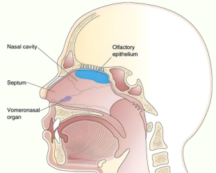
Newsletter Vomeronasal Organ (VNO) What is Vomeronasal Organ? While the human body is filled with intricate structures and organs, there are some that often remain hidden from the limelight. One such organ is the vomeronasal organ (VNO). Nestled deep within the nasal cavity, the Vomeronasal Organ plays a pivotal role in chemical communication and the perception of pheromones. In this article, we delve into the fascinating world of the vomeronasal organ, exploring its anatomy, function, and the ongoing debates surrounding its significance in humans. Anatomy of the Vomeronasal Organ The vomeronasal organ is a chemosensory structure found in many vertebrates, including humans, though its presence and functionality are subject to debate. Located within the nasal cavity, the It is situated close to the septum, just above the incisive canal. It consists of two narrow ducts, called the vomeronasal ducts, which open into the nasal cavity through small openings known as vomeronasal pores. Function and Significance The primary function of the Vomeronasal Organ is to detect and process pheromones. Pheromones are chemical signals released by individuals of the same species, which can influence various physiological and behavioral responses in conspecifics. It is highly specialized for detecting these chemical signals, which are important for social and reproductive interactions in many animals. In non-human mammals, the this organ plays a crucial role in mediating various behaviors, such as sexual attraction, territorial marking, and maternal-infant bonding. It is involved in detecting pheromones related to reproductive status, dominance, and social communication. However, the existence and functionality of this organ in humans are subjects of ongoing scientific investigation and debate. Debates and Controversies The presence and functionality of the Vomeronasal Organ in humans have been a topic of controversy within the scientific community. Some researchers argue that while the Vomeronasal Organ develops during embryonic development, it regresses and becomes non-functional in humans, rendering it obsolete in adulthood. They propose that other olfactory structures, such as the main olfactory system, play a more significant role in chemical communication. On the other hand, proponents of the Vomeronasal Organ’s functionality in humans argue that it may still play a role, albeit a diminished one, in mediating subconscious responses to pheromones. They suggest that this organ might contribute to modulating social behaviors, such as mate selection and certain emotional responses. However, further research is needed to reach a definitive conclusion on the exact functionality of this organ in humans. Conclusion The vomeronasal organ, although a small and enigmatic structure within the human nasal cavity, holds great potential for unraveling the complexities of chemical communication and pheromonal perception. While its functionality in humans is a subject of ongoing debate, this organ remains an intriguing area of research. Continued scientific investigations into the Vomeronasal Organ’s anatomy, molecular biology, and evolutionary history will shed light on its true significance and potential impact on human behavior. Library Anatomy Anesthesiology Biochemistry Cardiology Dermatology Emergency Endocrinology ENT Examinations Forensic Med. Obs. & Gynae. Hematology Medicine Microbiology Nephrology Neurology Oncology Ophthalmology Orthopaedics Paediatrics Parasitology Pathology Pharmacology Physiology Psychiatry Pulmonology Radiology Rheumatology Surgery

Read More
Vomer bone

Vomer Bone

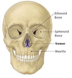
Newsletter Vomer Bone What is Vomer Bone? The vomer is a small bone of the facial skeleton (or viscerocranium). The vomer is one of the unpaired facial bones of the skull. This name is derived from the Latin word for a ploughshare and the shape of the bone. It is located in the midsagittal plane of the skull. Vomer bone forms the posteroinferior part of the nasal septum, with the superior part formed by the perpendicular plate of the ethmoid bone. This articulates with the sphenoid, the ethmoid, the left and right palatine bones, and the left and right maxillary bones. The name is derived from the Latin word for a ploughshare and the shape of the bone. Vomer bone has horizontal projecting superior expansions of bone called the ala of the vomer (wings) and a vertical perpendicular plate, which forms the main part of the bone. The vomer has two surfaces. On each surface, it presents a prominent oblique groove for the nasopalatine nerve and vessels called the nasopalatine groove. The isolated bone has four borders: anterior, posterior, superior, and inferior. The thickest border is the superior border which possesses a deep furrow between projecting alae, which fits the rostrum of the body of the sphenoid bone. Borders of Vomer Bone The anterior border is the longest and slopes downward and forward. Its upper half is fused with the perpendicular plate of the ethmoid; its lower half is grooved for the inferior margin of the septal cartilage of the nose. The posterior border is free of bony articulation, having no muscle attachments. It is concave, separates the choanae, and is thick and bifid above, thin below. The superior border, the thickest, presents a deep furrow, bounded on either side by a horizontal projecting expansion of bone – called the wing of vomer; the furrow receives the rostrum of the sphenoid, while the margins of the alae articulate with the vaginal processes of the medial pterygoid plates of the sphenoid behind, and with the sphenoidal processes of the palatine bones in front. The inferior border articulates with the crest formed by the maxillæ and palatine bones. Articulations of Vomer Bone The human vomer bone articulates with six bones: Two of the cranium, the sphenoid and ethmoid. Four of the face, two maxillae; and two palatine bones. The inferior border of the vomer articulates with the maxillae and the palatine bones. The superior border articulates with the sphenoid bone. The anterior border articulates with the perpendicular plate of the ethmoid bone and the nasal septal cartilage. The posterior border, however, is not attached to any bones, making it one of the few bones of the skull which does not fully articulate to other bones. This border separates the posterior nasal apertures or choanae. Vomer bone also articulates with the septal cartilage of the nose. Library Anatomy Anesthesiology Biochemistry Cardiology Dermatology Emergency Endocrinology ENT Examinations Forensic Med. Obs. & Gynae. Hematology Medicine Microbiology Nephrology Neurology Oncology Ophthalmology Orthopaedics Paediatrics Parasitology Pathology Pharmacology Physiology Psychiatry Pulmonology Radiology Rheumatology Surgery

Read More

ABC Nursing – Airway, Breathing, Circulation

Newsletter ABC in Nursing – Assessment & Priority ABC in Nursing – Airway, Breathing, Circulation The ABCs of nursing are a mnemonic that nurses can use to assist them remember the most important actions to take when prioritizing patient care. When prioritizing care, one must decide which needs must be attended to right once and which can wait until a later time due to less urgency. Maslow’s hierarchy of needs theory is utilized by nurses to prioritize patients and determine which patient needs to be seen first.  Airway, breathing, and circulation (ABC), which are physiological components required for the body to survive and contribute to a person’s level of health, are included in Maslow’s hierarchy of needs. A quick evaluation of life-threatening situations in terms of priority can be made by observing ABCs.  A stands for airway assessment, which involves checking for airway obstruction, which can be detected by stridor, a changing voice sound, and “see-saw” respirations. B stands for breathing assessment and includes checking for cyanosis, using accessory muscles for breathing, and an irregular respiratory rate. C stands for circulation, looking for changes in the color of the hands and fingers, an unusual capillary refill time, and a drop in consciousness (LOC).  When making an initial assessment, take into account a quick “see, listen, and feel” observation. Moreover, any obstruction involving the ABCs needs to be treated immediately. It should be highlighted that patient needs linked to maintaining a patent airway are always priority in prioritizing test questions, and that the ABCs should always be taken into account. You will be pointed in the right direction by this. Rish Academy Medical Notes  AIRWAY ASSESSMENT Problems with the patient’s airways that prohibit them from breathing or getting enough oxygen are referred to as airway difficulties. The patient’s breathing airways become constricted, obstructed, or swollen as a result of these obstructions. Potential Causes are Food blockages, Throat swelling, Foreign objects, and Chocking hazards/small toys Change in Voice The top goal when examining a patient’s airway is to check for any potential obstructions. An indicator that the airway is patent is if the patient responds with a normal voice. If the patient is speaking normally, the airway may be clean. Air entry is reduced and frequently noisy in a partially blocked airway. There will be no air movement or sound at the mouth or nose and a totally blocked airway. “See-saw” Respirations When an airway is completely or partially blocked, “see-saw” respirations are a common breathing pattern. Contrary to how it should usually move, airway blockage causes the chest to expand during exhalation and contract during inhalation. Stridor A loud, high-pitched, and harsh respiratory sound is stridor, often known as wheezing. This may indicate some airway blockage. A frequent indicator of airway blockage is snoring. Click the button below to Download 570+ High-Yield Presentations in Emergencies, Orthopedics, Gynaecology & Obstetrics, Surgery, and Clinical Medicine Get LIFETIME Access to 570+ Medical Presenations Some slides from the presentations from our Emergency Medicine Presentations  BREATHING ASSESSMENT The focus of breathing problems is on respiratory and ventilation problems. Many problems revolve around oxygen utilization. Fundamentally, this refers to how well individuals get enough oxygen into their lungs and blood. It also emphasizes how well patients expel toxic CO2 and oxygen from their bodies. An example would be when a patient has respiratory problems, lung illness, infections, asthma, allergies, inflammation, etc. 12–20 breaths per minute is the normal respiratory rate. Adults typically breathe 12 to 20 times each minute on average. A rapid breathing rate is a sign that the patient might be becoming worse. Use of Accessory Muscles in Respiration Accessory muscles are additional muscles that can help increase the amount of air that is inspired. These muscles are used to increase the volume and rate of respiration. Patients may appear as though they are pausing for breath between words, as the effort to breathe becomes more and more difficult. In adults, an observation of abdominal breathing is also an indication that breathing is labored. Cyanosis Cyanosis, or blue staining of the skin and mucous membranes, is caused by insufficient oxygenation of the blood. Abdominal breathing and perspiration are two additional typical indicators of respiratory distress. When assessing patients breathing it’s essential to look for signs of breathing difficulty. Basically, you want to look for visual signs and listen for sounds that indicate potential breathing/respiratory distress. This includes color changes of the face/skin, grunting, nose flaring, wheezing, and supplemental body positioning due to oxygen issues. Finally, check the patient’s pulse oximetry to indicate abnormalities that cause/lead to respiratory issues. Abnormal blood oxygen levels may indicate problematic oxygen saturation of the blood. Consequently, shortness of breath, confusion, and restlessness are potential signs of hypoxemia (low blood oxygen). Get LIFETIME Access to Emergency Medicine Notes CIRCULATION ASSESSMENT Circulatory assessment involves patients dealing with insufficient blood circulation. Circulatory problems result from various complications including physical occlusions, blood clots, diabetes, obesity, anemia, hemoglobin issues, etc. Basically, poor blood circulation leads to reduced blood flow and this can cause individuals to experience physical pain, muscle cramps, numbness, and tingling/stinking pains. However, this may also cause strokes, heart attacks, or other severe complications in extreme cases. Color of Hands and Digits Inspecting the skin will give the healthcare provider clues about any circulatory problems. Inadequate oxygenation of the blood leads to cyanosis or bluish discoloration of the skin and mucous membranes. The temperature of the skin should also be noted. In this case, the peripheries will be cold.  Normal Capillary Refill Time: 2 Seconds Checking the patient’s capillary refill time (CRT) is the easiest way to check circulation. To check the capillary refill time (CRT), the patient’s hand should be at the level of their heart. The healthcare provider should apply cutaneous pressure for 5 seconds with enough pressure to cause blanching. Time how long it takes for the skin to return to the color of the surrounding skin after releasing the pressure. The normal value for CRT is usually < 2

Read More
skin tag on nipple cov

Skin Tag On Nipple

Newsletter Skin Tag On Nipple Skin tag on Nipple Skin tags on nipples are a common condition that can appear anywhere on the body. Skin tags can be unattractive, uncomfortable, or irritable even though they are mostly harmless and don’t need to be treated. Skin tags are small, soft, non-cancerous growths that frequently have a stalk. Although they are harmless, dealing with them might be difficult. It makes no difference if you develop one skin tag or several. If you don’t already have any skin tags, you’ll probably develop some throughout the course of your life. Skin tags are common around the nipple because they can develop everywhere on the body, including around the breast. These tags are unharmful skin growths that have no chance of developing into cancer. They develop as a result of friction created when skin creases rub up against one another. Skin tags have a thin, flexible fleshy stalk that connects them to the skin and can range in size from one millimeter to one centimeter. They may be unpleasant and painful in some areas, such as the nipple, even if they are worried about it. The causes, signs, and treatments for skin tags on the nipple will all be covered in this article. have any concerns about a skin tag on your nipple. Skin Tag on Nipple What are skin tags? Skin tags are tiny, benign growths that often appear on the neck, armpits, and groin—areas of the body where skin rubs against itself or clothing. They can range in size from the size of a tiny pinhead to a few millimeters in diameter and are frequently characterized as looking like miniature balloons or mushrooms. Skin tags can be flat or elevated and are often flesh- or somewhat darker-colored. What causes skin tags on the nipple? Skin tags are thought to form as a result of friction or rubbing between the skin and clothing or other skin surfaces, while their specific cause is unknown. Given that skin tags are more prevalent during pregnancy and in people with specific medical disorders like diabetes, hormonal shifts may also contribute to their growth. Breastfeeding can also result in skin tags on the nipple. Skin tags can form as a result of minor skin tears brought on by an infant latching onto the nipple. Get LIFETIME Access to Medical Notes What are the risks of having skin tag on nipple? When you wake up with a skin tag on your breast, it can be a frightening and disturbing experience, especially if it’s your first time. While being benign, nipple skin tags can nevertheless be troublesome since they might irritate your bra or tight-fitting clothing. Most women want it gone because it makes them hurt, unhappy, and ashamed, especially while they’re dating. Also, you need to exercise extra caution with anything that pertains to your nipple in order to prevent aggravating your skin tag. You risk laying flat on your skin tag if you don’t pay attention to your sleeping positions. For mothers of newborns, skin tags on the breast might be problematic. except when What are the factors responsible for skin tag on nipple You are well aware that skin tags grow and form in areas where there is friction. The growth and development of skin tags are controlled by a number of variables. The woman’s weight is one of these factors. Skin tags are more likely to develop in individuals who are obese because there are more skin creases to cause friction. Because friction is thought to be caused by skin-to-skin contact, there are numerous different ways for the skin to get tense and result in nipple skin tags, especially in women. In expecting mothers, nipple skin tags are not unusual. In most cases, skin tags are likely to grow in number. Due to the swelling and enlarged stomach, the breast and stomach may rub against one another and cause skin tags. The most common location for skin tags in the breast is therefore below the nipple rather than on it. Due to constant friction from the baby sucking on the nipple, skin tags can develop on the breast of nursing mothers. Because sucking skin tags irritates and worsens over time, which could lead to more issues for nursing mothers, sucking the nipple will eventually cease to be a good alternative for babies. Younger women may experience skin tags because of the breast pressing against the cloth. Friction can happen when the nipple rubs up against tightly fitting clothing or an uncomfortable bra. Skin tags can also develop as a result of nipple piercings. It is not a good idea to wear a bra over a pierced nipple, especially since the pierced portion of the nipple can rub against the piercing and the bra. It will aggravate the condition by hurting the nipple and growing skin tags. Click the button below to Download 570+ High-Yield Medical Presentations in Emergencies in Medicine, Orthopedics, Gynaecology & Obstetrics, Surgery, and Clinical Medicine Get LIFETIME Access to Medical Notes What are the symptoms of skin tags on the nipple? The majority of the time, skin tags on the nipple are harmless and have no symptoms. However, if they rub against clothing or other skin surfaces, they may be unattractive and uncomfortable or irritate the skin. Rarely, skin tags on the nipple may bleed or get infected, resulting in discomfort and swelling. How are skin tags on the nipple diagnosed? A medical expert can identify skin tags on the nipple through a physical examination. To rule out any more underlying diseases, a biopsy may occasionally be carried out. How are skin tags on the nipple treated? Generally speaking, skin tags on the nipple don’t need to be treated because they are benign and safe. But, there are a number of ways to get rid of them if they are making you uncomfortable or irritated. Skin tags can be removed by cauterization, which involves using a particular tool to burn the tag off.

Read More
buy-usmle-step-3-notes-medical-notes-rish-academy

Buy USMLE Step 3 Notes

Newsletter Buy USMLE Step 3 Notes Why do you need to buy USMLE Step 3 notes? USMLE Step 3 notes are amazing guiding tools to remember complex theories if you are a medical, dental, or nursing student. There are a lot of books on the internet and in your university library with deep theories with long paragraphs. Although everyone uses USMLE Step 3 books for reference, most of the students refer to short notes for their studies and during exams. Because short USMLE Step 3 notes are easy to understand and remember complex theories very easily.  But unfortunately, there are only very few platforms on the internet that offer USMLE Step 3 notes. To solve this global problem and to help thousands of medical students worldwide, we have created a lot of USMLE Step 3 notes and presentations and you all can use them on our website for completely free. But to download our USMLE Step 3 notes PDF files, we charge a small fee to cover our expenses.  Rish Academy Medical Notes Where can you buy USMLE Step 3 notes? To make it easy for customers, we have made a very simple SHOP PAGE  where you can easily find the required notes in several USMLE Step 3 subjects. Up to now, we have more than 570+ USMLE Step 3 notes in several subjects like Clinical Medicine, Emergencies, Gynecology, Obstetrics, Surgery, and Orthopedics. Go to the SHOP PAGE, select the notes, click buy, and enter your payment details to get Lifetime Access to our USMLE Step 3 notes. To make a more trustworthy service, we only use PayPal and Stripe in our payment gateways which are the two most popular and secure payment gateways worldwide.  Along with our notes, you can also buy our popular Pharmacology, Pathophysiology, and Medical Mnemonics eBooks to boost your knowledge.  Click the button below to Download 570+ High-Yield Medical Presentations in Emergencies in Medicine, Orthopedics, Gynaecology & Obstetrics, Surgery, and Clinical Medicine Get LIFETIME Access to Medical Notes Some slides from the presentations from our Medical Resources Library Where can you download free USMLE Step 3 notes? We also offer Free, comprehensive, easy-to-understand USMLE Step 3 notes covering all the medical specialties that you need to know at medical school. You can browse on our website to read online and download them for free. There are numerous free USMLE Step 3 presentations on our website that are easily accessible to our users.  Share this : [Sassy_Social_Share total_shares=”ON”] Library Anatomy Anesthesiology Biochemistry Cardiology Dermatology Emergency Endocrinology ENT Examinations Forensic Med. Obs. & Gynae. Hematology Medicine Microbiology Nephrology Neurology Oncology Ophthalmology Orthopaedics Paediatrics Parasitology Pathology Pharmacology Physiology Psychiatry Pulmonology Radiology Rheumatology Surgery

Read More
buy-usmle-step-2-notes-medical-notes-rish-academy

Buy USMLE Step 2 Notes

Newsletter Buy USMLE Step 2 Notes Why do you need to buy USMLE Step 2 notes? USMLE Step 2 notes are amazing guiding tools to remember complex theories if you are a medical, dental, or nursing student. There are a lot of books on the internet and in your university library with deep theories with long paragraphs. Although everyone uses USMLE Step 2 books for reference, most of the students refer to short notes for their studies and during exams. Because short USMLE Step 2 notes are easy to understand and remember complex theories very easily.  But unfortunately, there are only very few platforms on the internet that offer USMLE Step 2 notes. To solve this global problem and to help thousands of medical students worldwide, we have created a lot of USMLE Step 2 notes and presentations and you all can use them on our website for completely free. But to download our USMLE Step 2 notes PDF files, we charge a small fee to cover our expenses.  Rish Academy Medical Notes Where can you buy USMLE Step 2 notes? To make it easy for customers, we have made a very simple SHOP PAGE  where you can easily find the required notes in several USMLE Step 2 subjects. Up to now, we have more than 570+ USMLE Step 2 notes in several subjects like Clinical Medicine, Emergencies, Gynecology, Obstetrics, Surgery, and Orthopedics. Go to the SHOP PAGE, select the notes, click buy, and enter your payment details to get Lifetime Access to our USMLE Step 2 notes. To make a more trustworthy service, we only use PayPal and Stripe in our payment gateways which are the two most popular and secure payment gateways worldwide.  Along with our notes, you can also buy our popular Pharmacology, Pathophysiology, and Medical Mnemonics eBooks to boost your knowledge.  Click the button below to Download 570+ High-Yield Medical Presentations in Emergencies in Medicine, Orthopedics, Gynaecology & Obstetrics, Surgery, and Clinical Medicine Get LIFETIME Access to Medical Notes Some slides from the presentations from our Medical Resources Library Where can you download free USMLE Step 2 notes? We also offer Free, comprehensive, easy-to-understand USMLE Step 2 notes covering all the medical specialties that you need to know at medical school. You can browse on our website to read online and download them for free. There are numerous free USMLE Step 2 presentations on our website that are easily accessible to our users.  Share this : [Sassy_Social_Share total_shares=”ON”] Library Anatomy Anesthesiology Biochemistry Cardiology Dermatology Emergency Endocrinology ENT Examinations Forensic Med. Obs. & Gynae. Hematology Medicine Microbiology Nephrology Neurology Oncology Ophthalmology Orthopaedics Paediatrics Parasitology Pathology Pharmacology Physiology Psychiatry Pulmonology Radiology Rheumatology Surgery

Read More