Drawing Brachial Plexus?
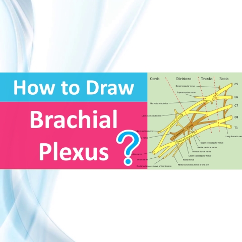
Newsletter Drawing Brachial Plexus There are several nerve plexuses in our body. The four main nerve plexuses are the cervical plexus, brachial plexus, lumbar plexus, and sacral plexus. The choroid plexus in the brain is a part of the central nervous system which consists of ventricles, capillaries, and ependymal cells. After reading this article, you will have a clear idea of drawing Brachial Plexus and understand how to remember them easily. The brachial plexus is a network of nerve fibers that supply the upper limb’s skin and muscle. It starts at the base of the neck, travels through the axilla, and ends in the upper extremities. The anterior rami (divisions) of cervical spinal nerves C5, C6, C7, and C8, as well as the first thoracic spinal neuron, T1, make up the plexus. The anatomy of the brachial plexus — its formation and anatomical course through the body – will be discussed in this article. Roots, trunks, divisions, cords, and branches are the five sections of the brachial plexus (a good mnemonic for this is Read That Damn Cadaver Book). There are no functional distinctions between these divisions; they are simply used to aid in the understanding of the brachial plexus. You can remember them with a picture shown above – ‘Tree and Swing’ Roots of tree – Five nerve roots are the five anterior primary rami of the spinal nerves Trunk of tree – Roots merge to form the trunks Trunk divides in tree – Each trunk then splits in two, to form six divisions Cords of the swing – These six divisions regroup to form the three cords or large fiber bundles Then most branches arise from cords (In above image, you have to remember the branches part separately) You can get access to our Medical Resources Library to get access to more than 500+ medical presentations and all other medical resources that will be helpful for your entire career. Get Lifetime Access to 500+ Medical Presentations and eBooks Follow these steps to draw Brachial Plexus 1) Roots The anterior and posterior ramus of each spinal nerve are then divided. The anterior rami of spinal nerves C5-T1 make up the roots of the brachial plexus (the posterior divisions innervate the skin and musculature of the intrinsic back muscles). These nerves emerge from the base of the neck, passing through the anterior and medial scalene muscles. The anterior and posterior ramus of each spinal nerve are then divided. The anterior rami of spinal nerves C5-T1 make up the roots of the brachial plexus (the posterior divisions innervate the skin and musculature of the intrinsic back muscles). These nerves emerge from the base of the neck, passing through the anterior and medial scalene muscles. When you draw the roots. first write nerve roots C5, C6, C7, C8 and T1 one below another. 2) Trunks At the base of the neck, the roots of the brachial plexus converge to form three trunks. These structures are named by their relative anatomical location: C5 and C6 roots combine to create a superior trunk.C7 continues up the middle trunk.C8 and T1 roots are found in the inferior trunk. When you draw, Join the C5 and C6 to form Superior Trunk/Upper Trunk C7 continues alone as Middle Trunk Join C8 & T1 to form Inferior Trunk/Lower Trunk 3) Divisions Within the posterior triangle of the neck, each trunk splits into two branches. One division goes anteriorly (toward the front of the body), while the other moves posteriorly (towards the back of the body). As a result, they’re referred to as the anterior and posterior divisions. Three anterior and three posterior nerve fibers are now present. These divisions pass via the axilla and out of the posterior triangle. They recombine into the brachial plexus cords. When you’re drawing, draw Anterior Division and Posterior Division from each trunk. You have to draw 6 divisions. 4) Cords Once the anterior and posterior divisions have entered the axilla, they join to form three cords, each called after the axillary artery that runs relative to it. The lateral cord is formed by: The anterior division of the superior trunk The anterior division of the middle trunk The posterior cord is formed by: The posterior division of the superior trunk The posterior division of the middle trunk The posterior division of the inferior trunk The medial cord is formed by: The anterior division of the inferior trunk. The cords give rise to the major branches of the brachial plexus. 5) Branches The three cords give rise to five primary branches in the axilla and the proximal portion of the upper limb. These neurons continue into the upper limb, where they supply innervation to the muscles and skin. These five nerves will be the focus of this section. Musculocutaneous Nerve Roots: C5, C6, C7. Motor Functions: Innervates the brachialis, biceps brachii, and coracobrachialis muscles. Sensory Functions: Gives off the lateral cutaneous branch of the forearm, which innervates the lateral half of the anterior forearm, and a small lateral portion of the posterior forearm. Axillary Nerve Roots: C5 and C6. Motor Functions: Innervates the deltoid muscles and teres minor. Sensory Functions: Gives off the superior lateral cutaneous nerve of the arm, which innervates the inferior region of the deltoid (“regimental badge area”). Median Nerve Roots: C6 – T1. (Also contains fibers from C5 in some individuals). Motor Functions: Innervates most of the flexor muscles in the forearm, the thenar muscles, and the two lateral lumbricals associated with the index and middle fingers. Sensory Functions: Gives off the palmar cutaneous branch, which innervates the lateral part of the palm, and the digital cutaneous branch, which innervates the lateral three and a half fingers on the anterior (palmar) surface of the hand. Radial Nerve Roots: C5 – T1. Motor Functions: Innervates the triceps brachii, and the muscles in the posterior compartment of the forearm (which are primarily, but not exclusively, extensors of the fingers and wrist). Sensory Functions: Innervates the posterior aspect of the arm and forearm,
Read More
